Table of Contents[Hide][Show]
In 2017, I had the opportunity to interview Dr. Philippa J. Johnson about her advanced imaging study for dogs with Degenerative Myelopathy. The Cornell researcher wanted to know if a cutting-edge MRI called, diffusion tensor imaging, could see lesions on a dog’s spine that’s associated with the disease.
Standard MRI (magnetic resonance imaging) isn’t able to detect the lesions caused by DM. The test, which is routinely given to dogs suspected of having the condition, is used to rule out other medical problems. It doesn’t diagnose degenerative myelopathy.
At the time, diffusion tensor imaging (DTI) had been successfully used to find microscopic changes in the spine of patients with ALS (Amyotrophic lateral sclerosis or Lou Gehrig’s disease). The information was extremely helpful to both patients and their doctors. It told them the stage of the disease and how quickly it was progressing.
ALS has many similarities to DM and the two diseases have been studied together often.
Dr. Johnson and her team hoped if they used DTI on dogs, the results would be equally successful.
If that were the case, dogs could get a confirmed diagnosis and veterinarians could see how far the disease had developed. It also meant that laser therapy and physical therapy could be started sooner. Both of these treatments have proven to slow DM.
As you can see, a lot was riding on Dr. Johnson’s study.
Well, after 4 years, the results were finally published in the Journal of Veterinary Internal Medicine. The news is very encouraging.
Before I share the results, here’s a quick overview of DM

Degenerative myelopathy is a progressive neurologic disease. It strips away the protective outer coating on the spinal cord that controls mobility. Dogs with the disease show signs of weakness in their hind legs that eventually turns to paralysis. And as time goes by, the front limbs and organs are affected as well.
DM strikes older dogs between the ages of 8-14. Victims can survive 2-3 years, but many pass away in 6-9 months.
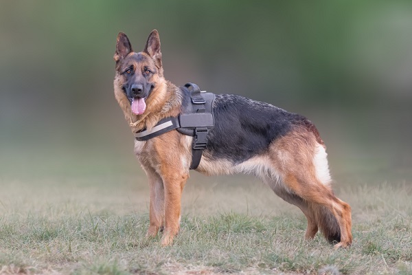
The disease is seen in a number of large breed dogs like German shepherds and Boxers and a few smaller breeds like Wheaton terriers and Corgis.
Click here to read the complete list of dog breeds prone to degenerative myelopathy.
Current methods for diagnosis
At the moment, dogs receive a diagnosis of DM based on their symptoms and by omission, which means other illnesses are ruled out.
The Orthopedic Foundation for Animals (OFA) has a DNA test, but it doesn’t diagnose degenerative myelopathy. It’s limited to telling pet owners if their dog is at-risk for developing the disease. The only way to positively confirm DM is through a necropsy (post mortem exam).
What is diffusion tensor imaging?

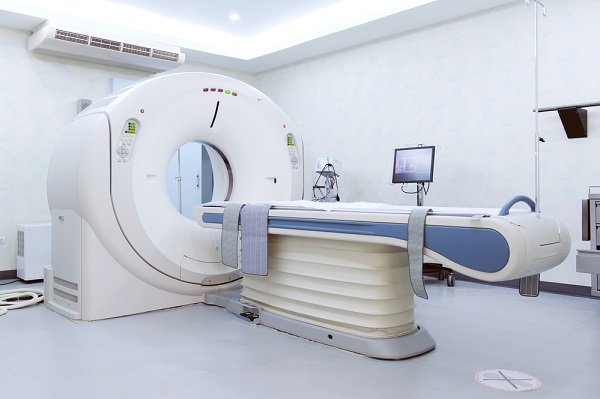
DTI is an advanced MRI technique that uses water molecules to generate images of the brain and spinal cord. Conventional MRIs use magnetic fields to create pictures, but DTI examines the direction and movement of water in the body.
The theory is that the movement of water isn’t equal. Obstacles like microscopic lesions, fibers and tissue redirect where the molecules go. This movement creates detailed images that can’t be seen anywhere else.
Results of the advanced imaging study for dogs with degenerative myelopathy

The study recruited 26 canines: 13 dogs with clinical signs of DM and 13 healthy dogs. All of the participants had to be at least 8 years-old. Dogs were represented by several different breeds. There was one Corgi, several German shepherds, several Boxers and 4 mixed breed dogs.
Most of the animals came from New York state, but two traveled from Michigan. The owner brought her dog with DM symptoms and the dog’s healthy housemate.
All of the subjects were examined one time at Cornell University. They each received a complete medical exam, blood work and a DTI procedure of their entire spine.
Pet owners had to agree that if their dog died during the time of the study, a post mortem exam would be done by the Cornell University pathologist.
The conclusion
The results confirmed that dogs suspected of having DM had significantly less strength in their spine, than the healthy dogs in the study. The strength was measured by a value called “fractional anistrophy” (FA) which evaluates the protective white matter in the spine. DM destroys that white matter. The decrease in FA showed up at each vertebra where a lesion was found.
It also showed the severity of the disease. Dogs with advanced forms of DM had the most decrease in FA values.
The overall conclusion is that FA has the potential to be a biomarker for the development of spinal cord lesions in degenerative myelopathy.

Get the Essential Guide
The Essential Guide of Products for Handicapped Dogs e-book is a labor of love for me. I wrote it to answer your most pressing questions about where to find the best products for your wheelchair dog. You’ll find products you didn’t know existed and each will improve your dog’s quality of life. Print a copy and keep it by your side.
Here are excerpts from the original interview with Dr. Johnson in 2017

I hope you take a minute to read through this interview. Dr. Johnson is an amazing and dedicated veterinarian and researcher. An example, is that in order to conduct her study on advanced imaging, she first had to get a master’s degree in human neuroimaging.
It meant leaving her position as a veterinary radiologist so she could learn about DTI technology.
I wanted to take the knowledge I learned and improve the diagnosis for dogs with degenerative myelopathy. I hope that will lead to understanding the disease better.
Dr. Philippa J. Johnson, Cornell University
The interview about the advanced imaging study for dogs with degenerative myelopathy.

Q: Explain an overview of the study.
The problem we are addressing is that the standard MRI doesn’t see the lesions in the spine that form with canine degenerative myelopathy. And the DNA test can’t predict the onset of clinical signs. Some dogs who receive test results that say they are “at risk” to develop DM, never show signs of the disease. The two current tests leave us with more questions than answers.
Diffusion tensor imaging for ALS patients has proven it can detect lesions in the spine. It confirms the condition and tells patients how far their disease has progressed. Our study will use the identical imaging technique with the goal of getting similar results in dogs.
Q: How will the ability to see spinal lesions help dogs?
We’ll be able to tell owners how bad the lesions are and what stage of the disease their dog has. If they are in the early stages, we can advise owners to enroll their dog in a physical therapy program or other rehabilitation.
Dogs will also be helped because veterinarians will have a tangible method for monitoring the results of new treatments. And we’ll be able to monitor dogs at risk, but not yet sick.

More of the interview
Q: If your theory is correct, will diffusion tensor imaging be readily available to the average pet owner with a sick dog?
Most universities are getting tensor advanced imaging. If we prove it to be useful for diagnosing pets, more private veterinary hospitals will add them. DTI is one of the fastest growing fields. It could potentially be used in the diagnosis of epilepsy and brain tumors in animals.
Q: Will DTI cost more?
It shouldn’t be more expensive because it will be part of a hospital’s MRI machine.
Interesting reading more about DM?
Degenerative Myelopathy: What Pet Owners Should Know – Click here
Laser Therapy Prolongs the Lives of Degenerative Myelopathy Dogs – Click here


Leave a Reply